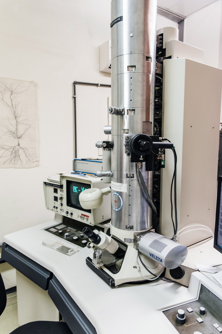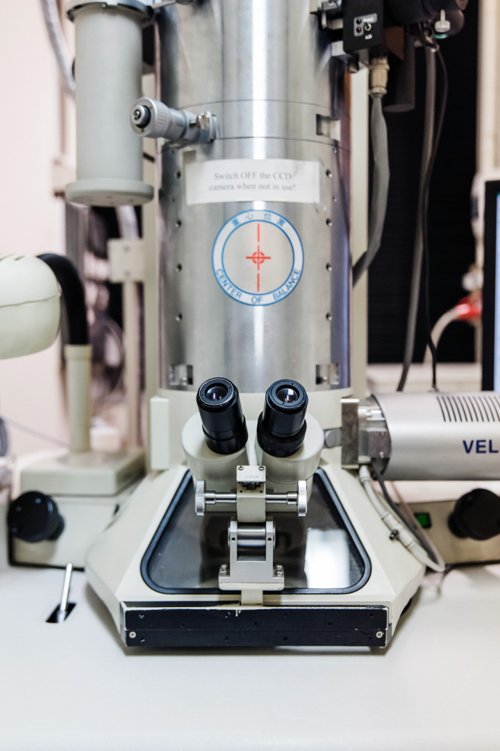
Electron Microscopy
Ultrastructural organization of synaptic connections is essential to understand the communication between neurons. This is especially true in the thalamus which displays the highest diversity of axon terminals in the brain. Both glutamatergic and GABAergic afferent terminals form morphologically and functionally distinct types of connections with thalamocortical cells.
These inputs differ in:
· how many synapses an individual axon terminals for
· which domains of their targets they contac
· what input pattern they conve
· what is their short term dynamic
· what is their impact on the behavio
· how they are combined with other types of afferents on thalamocortical cells
Since the ultrastructure of afferent terminals largely define their functions, clear functional predictions can be made as tested by physiological experiments just by studying thalamic inputs at the electron microscopic levels.
We made the following key discoveries using this method:
1). Thalamus receives the largest, most complex, multisynaptic GABAergic terminals in the brain from several subcortical sources. These inputs are conserved between rodents and primates, exert specialized, non-depressing inhibition and have a profound impact on sensory transmission, behavior and global brain activity via the thalamus.
Ø Barthó et al., 2002 - Multisynaptic GABAergic inputs from zona incerta
Ø Bokor et al., 2005 – Multisynaptic GABAergic inputs from the anterior pretectal nucleus
Ø Lavelle et al., 2005 – Effect of multisynaptic GABAergic inputs on sensory transmission
Ø Wanaberbecq et al., 2008 – Synaptic properties of multisynaptic vs unisynaptic GABAergic inputs
Ø Bodor et al., 2008 – Comparison of rodent and primate multisynaptic GABAergic inputs
Ø Giber et al., 2015 – Structure and function of multisynaptic GABAergic inputs from the brainstem
Ø Halassa & Acsady 2016 – Review of multisynaptic vs unisynaptic GABAergic communication in the thalamus
2). Individual thalamocortical cells can receive complex multisynaptic excitatory terminals, which can drive their output, from two different sources (cortex and brainstem). As a consequence these neurons do not act as “relay” neurons, rather they integrate the activity of two distinct brain regions.
Ø Groh, Bokor et al., 2014 – Integration of cortical and subcortical driver inputs by thalamocortical cells
Primate thalamus receives a large variety of excitatory input types from both cortical and subcortical sources. Parcellation of primate thalamus based on excitatory input types and sources revealed that the classical “sensory” synaptic organization is present only in the minority of thalamus. Synaptic organization of the majority of the thalamus is different. E.g. large territories are dedicated entirely to cortico-cortical communication via the thalamus whereas in other regions large multisynaptic excitatory inputs are missing entirely.
Rovo et al. , 2012 - Mapping macaque thalamus based on its excitatory afferents.