


Electron microscopy
Sunday, 20 March, 2022
Ultrastructural detection of synapses between kisspeptin neurons of the human hypothalamus
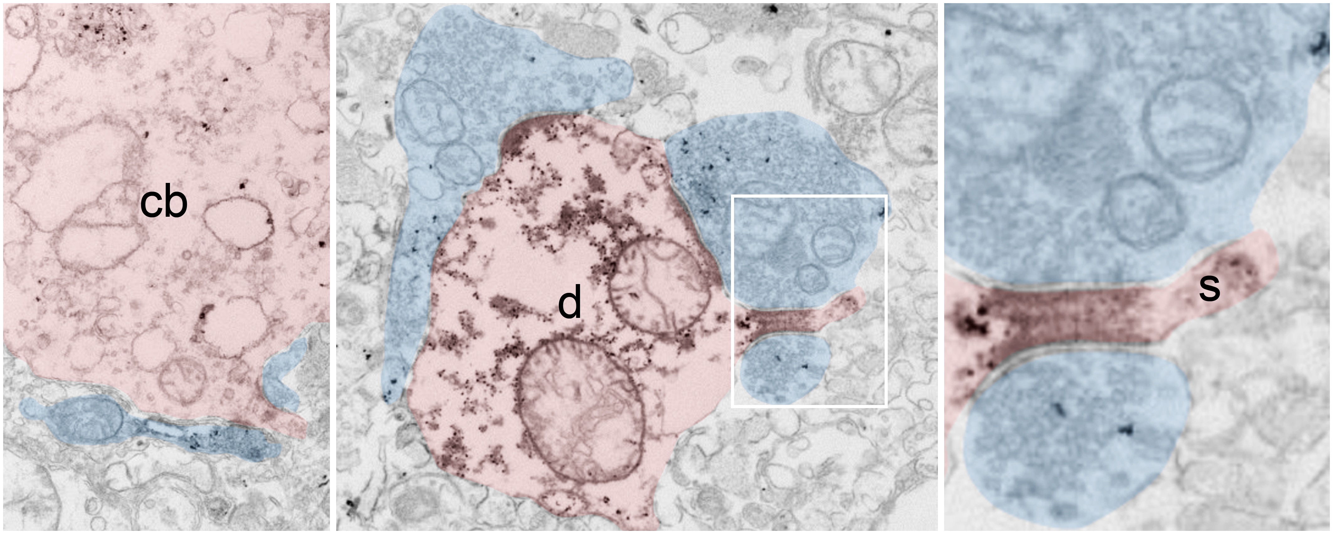
Short post mortem-time immersion-fixed hypothalami or perfusion-fixed samples provided by the Human Brain Laboratory of the Institute are compatible with immunoelectron microscopy. Figures reveal kisspeptin-immunoreactive axons (semi-transparent blue) establishing asymmetric synapses with kisspeptin-immunoreactive (semi-transparent red) cell bodies (cb), dendrites (d) and dendritic spines (s) in the human infundibular nucleus.
Takács et al., Brain Struct. Funct., 223 (5) (2018), 2143-2156