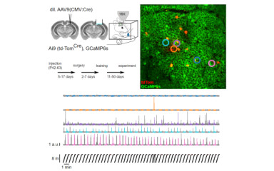
Answering many scientific questions requires the simultaneous examination of the activity of numerous neurons. Our group is able to monitor the activity of more than 500 cells simultaneously with two-photon (2p) Ca2+ imaging.
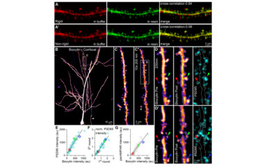
SDS-FRL is a modern method of electron microscopic immunolocalization with superior sensitivity and resolution compared to conventional pre- and post-embedding immunolocalization techniques. The technique provides a two-dimensional view of the neuronal cell surface, which facilitates the measurement of cell surface areas, and the antibodies do not need to diffuse into the tissue during the immunoreaction, making it ideal for quantitative analyses. Gold particles of different sizes can be attached to antibodies that specifically label the membrane protein of interest, allowing the quantitative investigation of their cell surface distribution and their enrichment in different subcellular compartments with nanometer precision. The technique is also ideal to study the intra-synaptic distribution of synaptic proteins.