


A small step closer to understanding schizophrenia
Schizophrenia is a serious neurological disorder that affects tens of millions of people and requires lifelong medication and psychosocial support. Effective, targeted treatment requires an understanding of the pathological processes involved, which in turn requires mapping the changes occurring in the neural networks of the brain areas affected by the disease at the cellular, synaptic, and molecular levels. The Nusser group has developed a groundbreaking, high-resolution technique that allows the study of synaptic proteins at the level of individual synapses in human brain tissue. Using this method, they discovered a selective molecular change (decreased NMDA receptor density) in a specific subset of excitatory synapses in schizophrenic patients.
It is known that the dorsolateral prefrontal cortex (DLPC) plays a particularly important role in the development of cognitive disorders in the pathophysiology of schizophrenia. Genetic and transcriptomic data suggest a disturbance in excitatory, glutamatergic synaptic transmission. Since mRNA levels do not always proportionally reflect the amount of proteins produced, changes in synaptic function must be examined at the protein level. However, previous protein measurements (e.g., using Western blot) only provided information about the average protein content of the entire cerebral cortex. High-resolution microscopic data revealing the distribution of synaptic proteins have so far come almost exclusively from rodents, as it is still a major technical challenge to detect and recognize molecules embedded in the dense protein network of synapses in postmortem fixed human brain tissue remains a serious technical challenge to this day. The Nusser group's methodological development has brought about a breakthrough in this area.
The aim of the group's research was to explore the molecular composition of excitatory synapses at the level of individual synapses in postmortem human control and schizophrenic dorsolateral prefrontal cortex. The samples for the studies were provided by the KOKI Human Brain Tissue Laboratory.
Andrea Lőrincz, the first author of the article: "Our research group has successfully optimized a highly sensitive post-embedding immunofluorescence labeling method for human tissue. Using our method, we were able to obtain quantitative data on the amount and distribution of synaptic proteins from thin (200-400 nm) sections embedded in synthetic resin using confocal and STED super-resolution microscopy with a resolution of up to 40 nm. In strongly fixed human tissue, we made synaptic proteins accessible to antibodies using a microwave antigen retrieval step. A major advantage of our method is that, after removing each antibody, dozens of different proteins can be labeled in the same synapses in several consecutive steps!"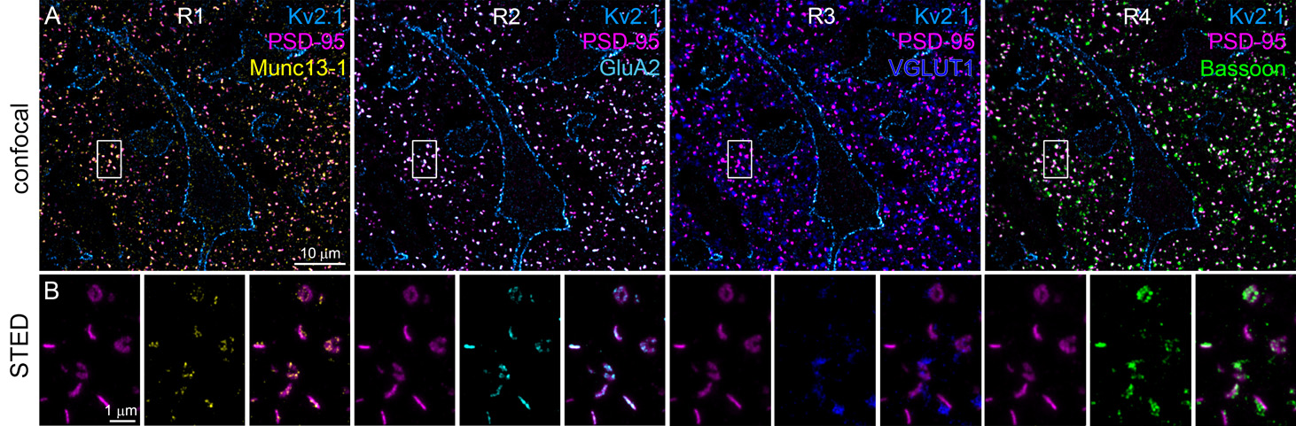
Confocal and STED (Stimulated Emission Depletion) images showing multiplexed immunofluorescent labeling of a 300 nm thin resin-embedded section obtained from a control human prefrontal cortex. Four rounds (R1-R4) of triple labelings were sequentially performed on the same set of synapses to identify the precise spatial organization of multiple synaptic proteins.
The overall stability of excitatory synapses was a surprising result. There was no detectable difference in the density and size of excitatory synapses in the upper cortical layers of control and schizophrenic patients, and the amount of AMPA and NMDA receptors (GluA2, GluN1, GluN2B subunits) and presynaptic molecules (Bassoon and Munc13-1) were also similar between the two groups. Even the organization of neurotransmitter release sites remained intact in most synapses, suggesting that the synaptic disturbance associated with schizophrenia may not be global but limited to specific neuron types. The examination of parvalbumin-expressing (PV+) inhibitory interneurons, cells known from the literature to be affected in schizophrenia, confirmed previous findings that in schizophrenic samples, the PV protein level in the cell bodies of PV+ inhibitory interneurons in schizophrenic samples is approximately 30% lower than in control samples. Examination of excitatory synapses directly on the dendrites of PV+ interneurons showed that although the amount of synaptic AMPA receptors was similar between the control and schizophrenic groups, the synapses of schizophrenic patients contained fewer NMDA receptors.
Andrea Lőrincz: "Our results suggest that the development of schizophrenia pathophysiology involves changes in the molecular composition of excitatory synapses in certain cell types, rather than a comprehensive disruption of all excitatory synapses in the prefrontal cortex.
PV+ interneurons play a key role in regulating network oscillations and cognitive functions. The reduced NMDA receptor density observed in the excitatory inputs to these interneurons suggests that the excitability and plasticity of PV+ cells may be impaired in schizophrenia. This may alter the balance between excitatory and inhibitory systems, contributing to the cognitive decline observed in schizophrenia. We hope that our method will promote the exploration of the role of molecules that may be decisive in the development and course of schizophrenia and other neurological and psychiatric disorders."
Lorincz A, Ashaber M, Nusser Z :Altered Molecular Composition of a Specific Subset of Prefrontal Cortical Excitatory Synapses in Schizophrenia. J Neurosci. 2025 Sep 17;45(38):e0645252025. doi: 10.1523/JNEUROSCI.0645-25.2025. PMID: 40829937; PMCID: PMC12444913.