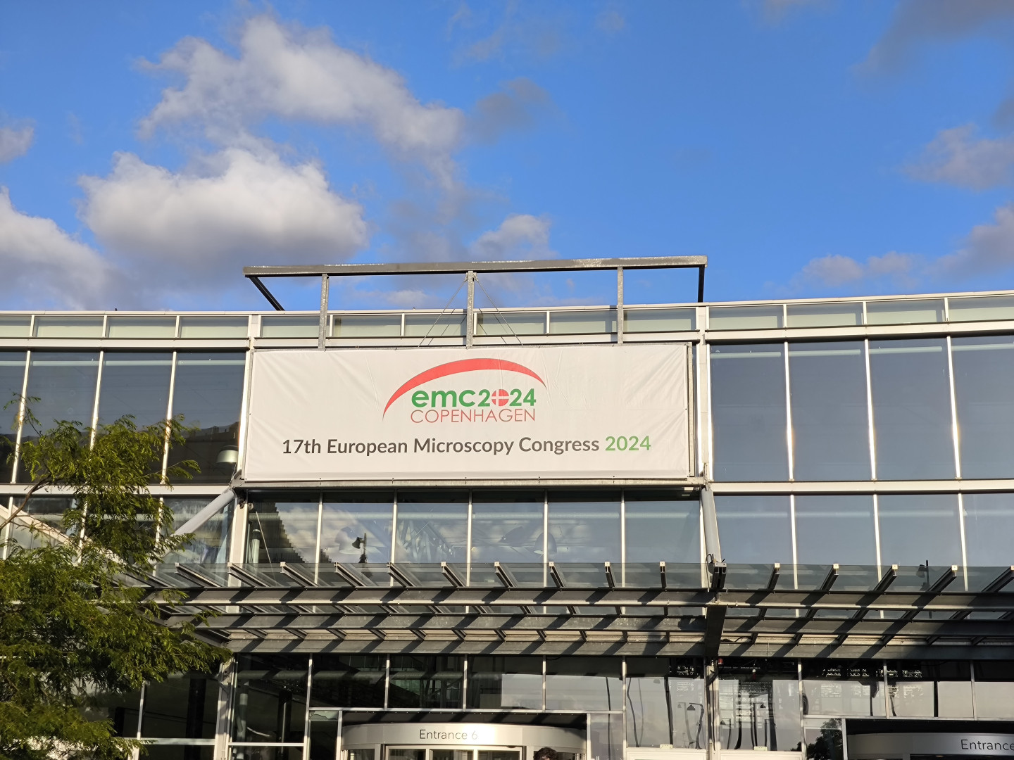
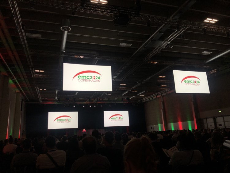
Our microscopists and the European Microscopy Conference in Copenhagen (EMC2024)
Copenhagen not only provided an excellent venue, but also a wonderful summer weather during the last week of August for the 2500 participants of EMC204, the largest meeting of microscopists in Europe. The Hungarian Microscopy Society was represented by Techoorg Linda and eight microscopists, including four colleagues from KOKI.
The microscopy conference in Lyon in 2016 was voted by the representatives of microscope societies from European countries not to be won by Barcelona on the Mediterranean coast or by Basel, which guarantees excellent organization (the advantages of Brno and Maastricht are not discussed here), but by Copenhagen, with its much less pleasant climate and even more expensive than the former.
Then COVID overruled everything, leaving only Copenhagen as the venue for the next European conference, as decided by the EMS board. To ensure that all the hard work invested so far would not go to waste, the right to host 2024 was awarded to Copenhagen without a tender. And the city, perhaps as a thank you, waited with the bright, pleasant summer days that are a rarity here.
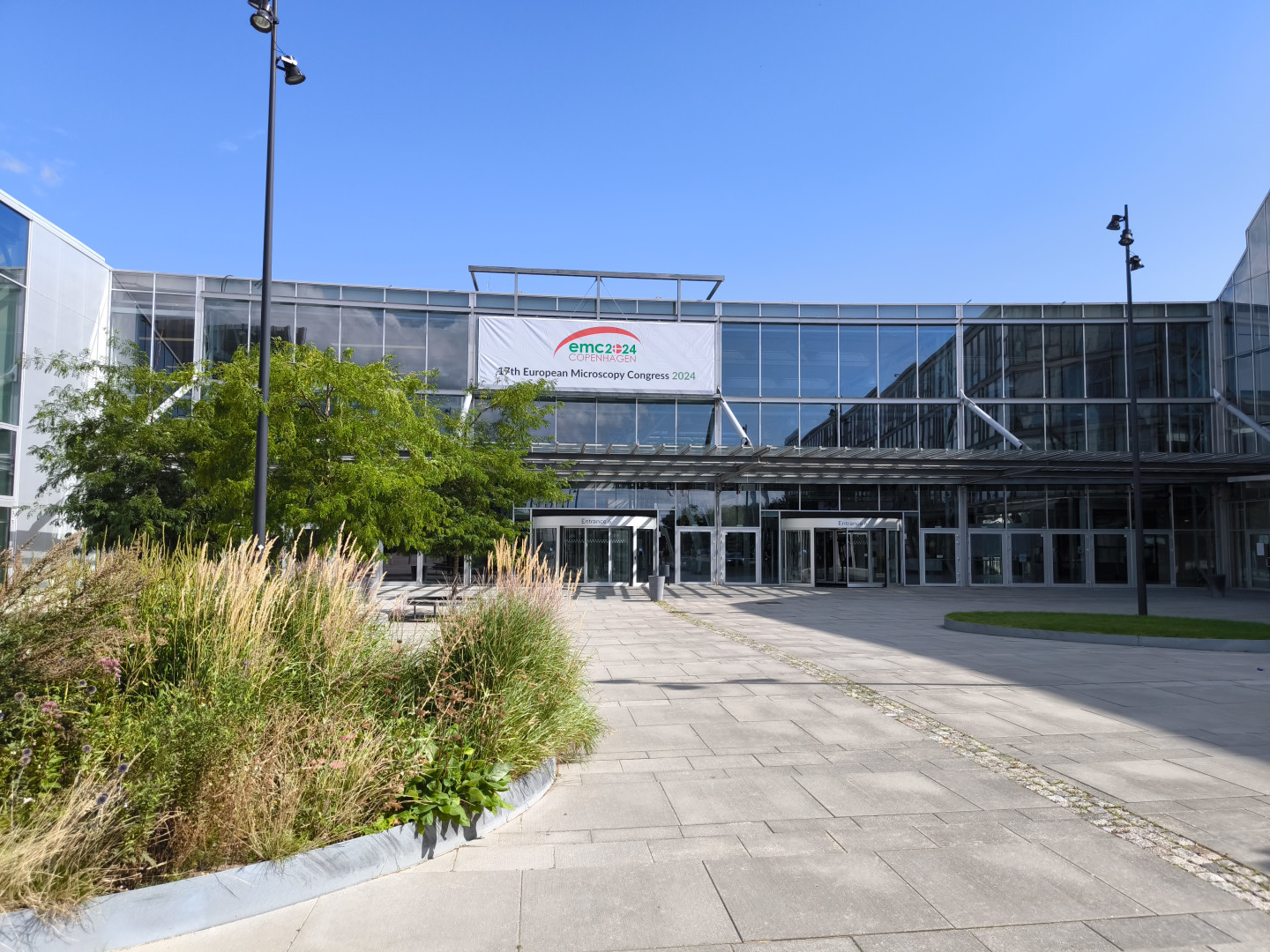
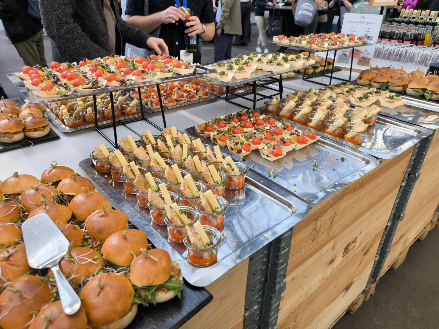
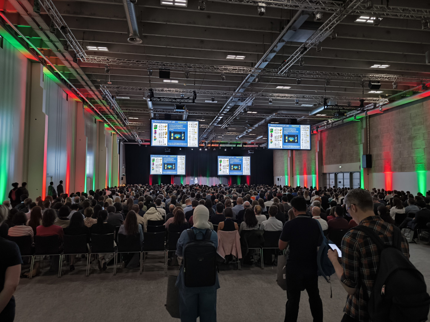
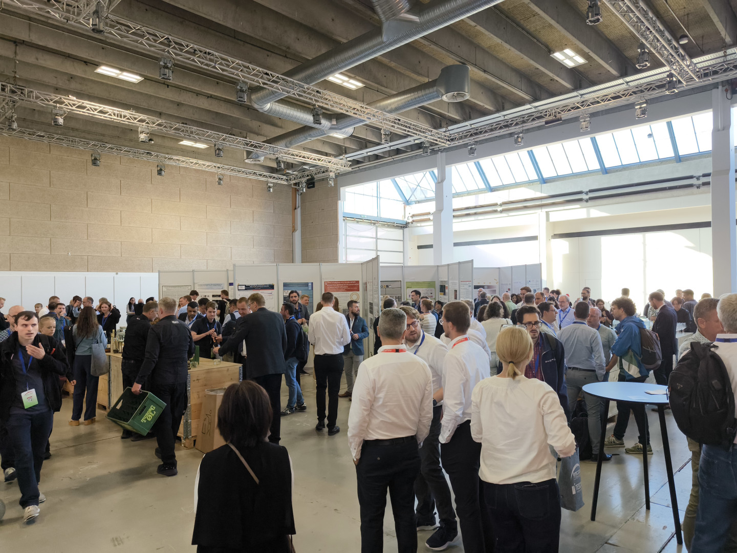
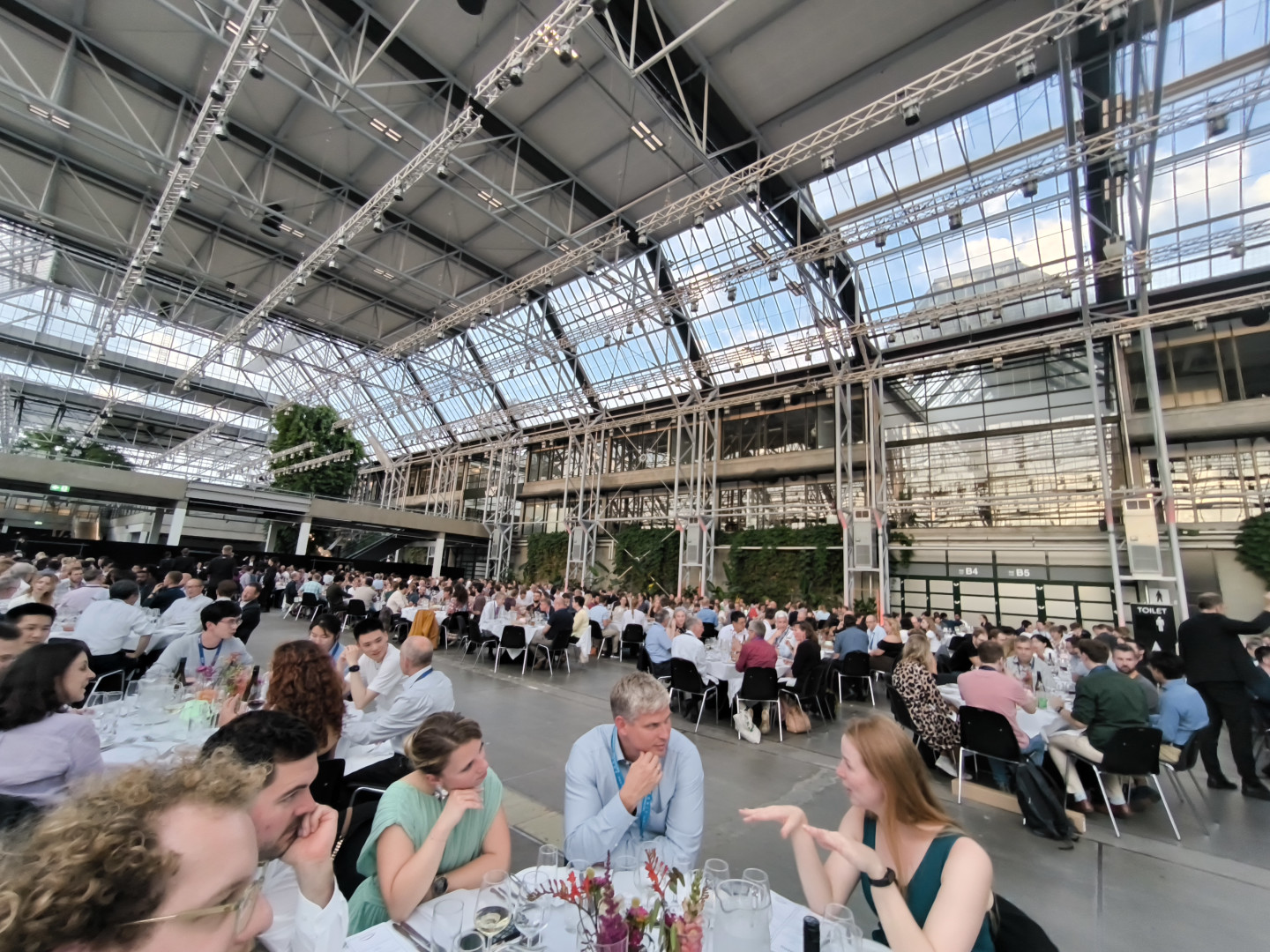
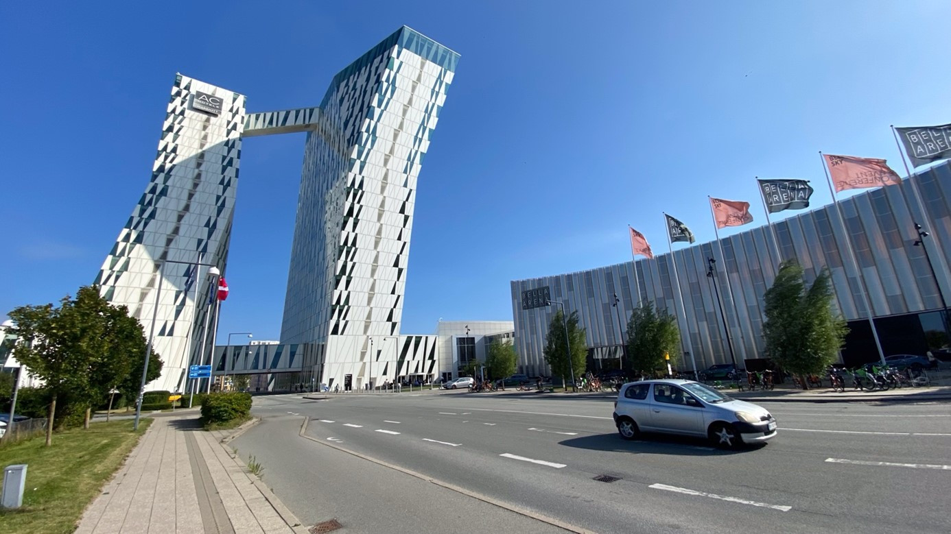
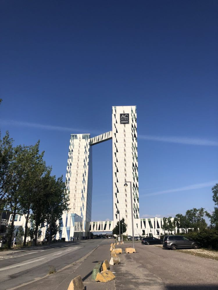
The conference venue, the Bella Center, was easily accessible from both the airport and the city center. Even though two conferences were going on simultaneously, the participants did not even disturb each other. The 2,500 registered EMC participants never felt lost in the crowd. The plenary sessions had a large hall with a capacity of at least 2,500 people, and the five parallel sessions had rooms for 3,500 to 500 people, with access from a large lobby, where the companies' exhibition stands and posters were located, tables offering iced water, hot tea/coffee, and a delicious seed mix all day long, and even more islands for relaxing on bean bags, eating, reading the program and, even more, chatting and meeting fellow microscopists from elsewhere.
It has long been common practice to organize workshops for a small number of people on a specific topic before or after the opening of the official program of major conferences. This was also the case in the Copenhagen program. The events, which were designed for 16 to 60 people, depending on the topic, included lectures and practical sessions and were held at the University of Copenhagen or one of the buildings of the University of Technology.
Before the official opening, Zsu Környei had already attended a workshop on the use of artificial intelligence in microscopy, where he admittedly also had to "hang on" to follow the speakers.
As befits a senior researcher, he was diligently taking notes throughout the event, particularly with a view to what could be tried at home, what could help solve current problems, and collecting resources where they could be accessed on the internet. He brought home a lot of useful information, which he has already shared with his group mates and made available to others.
The presentations at the conference focused on the diversity of technologies the possibilities of testing and the significant expansion of the range of possibilities.
- Behind all the acronyms lie exciting technologies, expanding the scope of testing. For example, Hybrid Unmixing (HyU) technologies for the detection of endogenous fluorophores (e.g. retinoids, NADH), well-penetrating so-called "HyU" technologies for the convincingly uniform staining of deep tissue layers, and the use of a variety of new technologies for the detection of endogenous fluorophores (e.g. retinoids, NADH). "In situ Host-Guest Chemistry (INSIGHT), DNA-printing techniques for single molecule detection, fluorescence microscopy with up to angstrom resolution (RESI), or ultrastructural expansion microscopy (U-ExM), which can be used for confocal microscopy. But then there are the advances in light plane microscopy, the multi-light sheet (RESOLFT), then the two-photon super-resolution microscopy (Two photon line-scanning structured illumination microscopy; LIL-SIM)... It is not easy to list the new techniques!
- Do you have any critical observations or comments to consider?
- I think that today some people are still lost in the glamour of technology, and biological relevance is more often left behind. However, there is no way around it, we have to keep up with technological developments, keep track of developments, and keep them constantly communicated to researchers and encourage them to use them. In addition, data analysis needs to be improved to ensure that as much information as possible can be extracted from the large amounts of data collected using the latest methods.
The head of the KOKI Light Microscopy Centre, Pál Vági, came to Copenhagen with his wife and son, three of them, via Norway.
VP
- I have been here many times, but never for professional purposes. As has happened at other big conferences, I got lost in the program on the first day.
I felt that I would not be able to fill the time, because I did not want to deal with electron microscopy topics there, and they were predominant. But then the picture cleared up and I always managed to sit in on sessions I could enjoy.
- What experiences have you had?
- Based on the lectures I have heard, it seems to me that correlative microscopy, where a series of different imaging modalities or methods are combined in the workflow with microscopes of disparate resolutions (light microscopes, electron microscopes) to analyze the same sample, is still a struggling genre. I did not feel that there had been any significant progress since the beginning.
- Yet there was more than one session on this topic! But were there any that were a pleasant surprise?
- Tuesday was the session that "really hit the spot", especially as the method discussed, expansion microscopy, started to engage me this summer.
We heard a fascinating talk by a Swiss researcher who has changed from a cryo-electron microscopist and tomography researcher to an expansion microscopist. He has solved almost every structure with a light microscope, even structures that electron microscopists today only dream of!
The origin of the presentation was a transmission electron microscope image of Professor Paul Röhlich's centriole. It was astonishing to see that this tiny structure, which until now could only be examined with the resolution provided by the electron microscope, and many others like it, can now be investigated by remaining in the realm of light microscopy. The number of markings is almost unlimited, the images are colorful and the whole process is free from the pains of electron microscopy sample preparation. The lecture group alone would have made the trip worthwhile.
In my experience, real microscopists speak similarly about microscopes, methods, and procedures close to their hearts. The subject itself distinguishes them from non-microscopists but makes them similar to everyone equally passionate about what they do.
- And what about the exhibitions of microscope companies?
- I spoke to three exhibitors, which was useful in the run-up to the microscope purchases expected at the end of the year. I was able to get to know a light-sheet microscope (miltenyibiotec.com), I talked to an exhibitor from a laser company and I learned that even a strange idea - two microscope systems with completely different illumination, both of which give excellent performance - can become a viable product if the idea is born in the right country (chipnano.com).
- Then it's worth visiting besides Northern Europe, a conference like this!
- Definitely! But it was also worth seeing smiling Copenhagen. We did great family activities in the empty runs, and it was hard to head back home, back into the sweltering heat.
And finally, what does someone who has attended a major microscopy conference for the first time have to say?
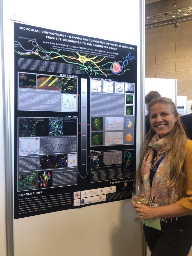
Dóra Anett Máté-Schwarz, aka Netti, a member of Ádám Dénes' group, who is doing her PhD work under the supervision of Csaba Cserép and Ádám.
- We heard high-quality presentations from many branches of the microscopy field, from physical principles to technical developments and life science applications. It was exciting to learn about topics specific to my discipline but also about completely different fields.
- What would you highlight?
- There were several presentations on the topic of correlation. This meant a special benefit for me. I also gained some ideas and techniques that could be useful for our experiments. I would mention as an example the inspiring presentation given by Jemima Burden. She shared her experiences with correlation methods, their importance, and their difficulties. The presentations showed a lot of wonderful 3D reconstructions, but they still took a long time and a lot of effort to create, as the speakers said. And the accuracy of automated software still does not approach human precision!
- You also presented a poster!
- Indeed. My poster presented the topic of my talk at this year's Hungarian Microscope Society conference and was extended with my new results. Despite my previous experience, I got mainly methodological questions and advice.
- How would you sum up your experience?
- Interestingly, electron microscopy methods and techniques were more prominent at this conference, with less discussion of light microscopy techniques. True, even after the few light microscopy methods most commonly used in correlative microscopy, the list of electron microscopy (EM) options is longer. Transmission EM (TEM) and tomography (ET), cryo-EM, volumetric EM (SBF-SEM, FIB-SEM, array tomography, etc.). Also increasingly popular is volumetric electron microscopy (vEM), which has been developed to visualize the ultrastructure of cells and tissues in 3D and at volume scales from μm to mm and at high resolution.
For 3D imaging of serial sections, ssTEM is used, ssET for multislice tomograms, for scanning EM (SEM) serial sections can be imaged by array tomography (AT), and the removal of block surface layers after SEM imaging is performed either by a focused ion beam (FIB-SEM) or by the built-in diamond knife (SBF-SEM). There were too many procedures and acronyms, and my list is incomplete!
In conclusion, I can say that the scientific standard of the conference was high. Although the information app was far from perfect, and I could imagine a better solution for the catering, overall it was useful, interesting, and exciting. I gained a lot of experience and it is the most important.