





"Transcriptome profiling"
The work of our institute’s Reproductive Neurobiology Research Group was published in the August 2026 issue of Nature Communications. Group leader Erik Hrabovszky, who directed and organized the work, is the last author of the article, and Szabolcs Takács, a member of his group and the first author of the publication, answers our questions.
- Congratulations on your Nature Communications paper and on being the first author. Please tell us about the work!
- It might be worth starting with DNA, which everyone is familiar with, or, more precisely, the genome, which is the totality of the hereditary information encoded in the DNA of a living organism’s cells. The entire population of RNA molecules transcribed from this at any given time is called the transcriptome.
The structure of a single DNA or RNA strand, or the proteins translated from it, is determined using sequencing methods. Transcriptomics is the high-throughput analysis of the transcriptome in a cell, cell type, tissue, or organ. High-throughput sequencing—also known as next-generation sequencing—has become widespread in recent years and is capable of determining the structure of thousands of molecules simultaneously.
- While we’re on the subject of defining terms, what is neuroendocrinology?
- Neuroendocrinology is a branch of the vast field of neuroscience that studies the interaction between the nervous system and the endocrine system, including the neural and even molecular underpinnings of the diverse neuroendocrine conditions and disorders that affect so many people. However, there is less information available about this important research—such as that on neurodegeneration—than there is about other topics.
- Your group’s findings have been published one after another in the top endocrinology journals in recent years, but why did you choose the NatComm this time?
- Although the topic of this article is neuroendocrinology, the method we developed has a much broader range of applications. We’re pleased that our study was published in a journal whose readership extends beyond the narrow field of neuroendocrinology. We hope that other researchers will also make use of the data and the method itself. The best possible outcome would be if one of the molecular characteristics we’ve now uncovered were to later take on functional or even pharmacological significance.
- And that brings us to my first “real” question! Why did you need to develop this method, and why is it necessary to study human neurons directly?
- Even in so-called basic research, the ultimate goal is quite rarely to understand laboratory animals at the molecular level. From our previous work, we are well aware of species-specific characteristics in neuroendocrine regulation, which is another reason why it’s important to demonstrate exactly how human cells differ from their rodent counterparts!
- What has prevented this so far?
- It’s not just one thing. Even in the case of animal experiments, a very rigorous ethical review process is required before a series of experiments can begin, and human tissue samples are typically obtained only from tissues removed during specific surgeries. And even then, only with authorization granted after an even stricter review process. For our studies, we needed deep brain samples, and suitable surgical material is particularly rare in this area.
- What was the biggest obstacle to applying your IHC/LCM-Seq sequencing method—which you developed for rodents—to human tissue?
- The essence of our method is that, without genetic modification, we identify and collect selected cells using a protein identified by immunohistochemistry, and from these we generate a high-depth transcriptomic profile. We had to adapt and then validate this method for postmortem human tissue. It was neither quick nor easy, but we succeeded in the end.
This workflow and the validation of the method have now been published in the Nature Communications.
- How does the new procedure applied to postmortem human brain tissue differ from the one used on rodents?
- In addition to having to optimize the fixation conditions, both the immunohistochemical processing and the laser microdissection are more complex and pose a greater challenge with these samples than with tissues from laboratory animals.
- Are there any elements or steps in the new procedure that were not used in the previous one?
- The incorporation of epitope mapping marked a new step, when we treated human tissue sections at approximately 80 °C. It was surprising that the RNA-protecting additives remained effective even at this high temperature. When we were able to extract RNA of sufficient quality even after treatment, it became clear to us that the procedure was viable for human tissue as well.
- When you saw or read the results, what was the most interesting or surprising thing for you?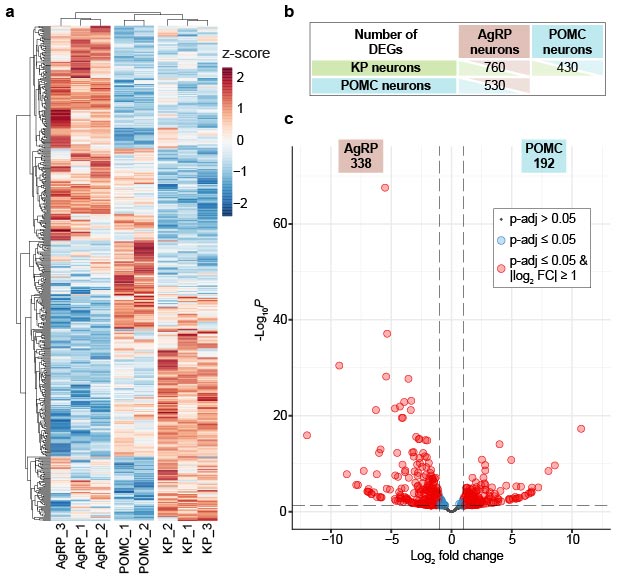
- Perhaps what surprised us most was the richness of the molecular repertoire of the cells we studied. We found a wide variety of neuropeptides and receptors, with marked human–rodent differences in many cases. A good example of this is the distribution of Substance P and CART peptides, which we confirmed with immunohistochemistry. This technique verified the presence of the proteins following the transcriptomic data. CART, for example, is strongly expressed in some human AgRP and kisspeptin neurons, whereas in rodents it is primarily associated with the POMC system.
- What might be the significance of these findings for the human body and, eventually, for medicine?
- By understanding which receptors and signaling pathways are used, for example, by the AgRP and POMC neurons that regulate human food intake, we can better assess which hormonal and metabolic signals or drugs might influence the function of these two cell types. One of the greatest contributions of our work is the mapping of the receptor landscape, which can also identify potential drug targets suitable for treating obesity, for example. Human data are also needed to confirm whether mechanisms that appear promising in animal models are relevant in humans.
The active ingredient in Ozempic—originally used to treat diabetes but now touted as a weight-loss miracle drug—stimulates GLP-1 receptors. We have successfully detected such receptors in both POMC and AgRP neurons. In addition, numerous other receptors could form the basis for promising future strategies in the fight against obesity.
- But these are still observations made during basic research . . .
- That’s true. Moreover, the presence of a receptor’s mRNA does not yet prove either its functional role or that it would be a suitable drug target. However, the data can identify candidates worthy of further investigation and help us rethink certain concepts derived from animal experiments. Using our method, we can generate high-depth profiles by sequencing hundreds of pre-identified nerve cells or other cell types, and we can reliably detect even the transcriptomes of rare receptors or other molecules present in small quantities.
- In other words, you’ve succeeded in significantly broadening the applicability of your earlier methodological developments. The inevitable question is: who was involved in the work?
- Erik Hrabovszky led the development. We began the experiments with Katalin Skrapits, Éva Rumpler, and Balázs Göcz, and we continued working primarily with Kata on the human application. Miklós Sárvári compiled the RNA-based molecular data and sequencing libraries; we received the human samples from pathologists at Semmelweis University, and the sequencing was carried out with the collaboration of our colleagues in Debrecen.
The supplementary data requested by Nature Communications also substantially strengthened the study. Dalma Biri and Éva Rumpler validated several transcriptomic observations through immunohistochemical analyses, while Barbara Göblyös did so using RNAscope. A regional comparison of POMC neurons was also conducted at this time.
- This phase is complete. What’s next?
- The published transcriptomic results were derived from male donors. The kisspeptin neurons in the region examined in our current experiments are not only central to the regulation of reproduction but also play a key role in the development of menopausal hot flashes. We hope that our experiments using samples from female donors will help shed light on these processes as well.